✓ US FDA 510(k) Cleared
✓ CE Certified

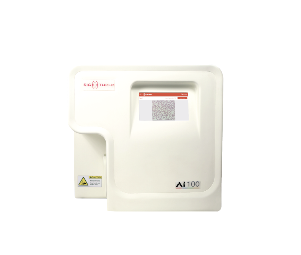
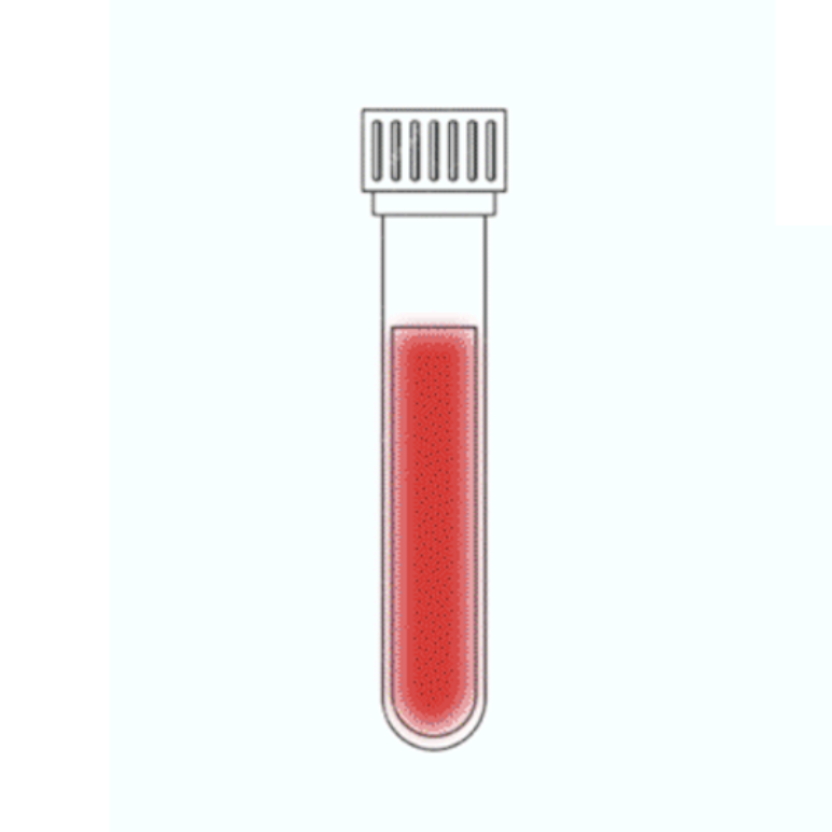
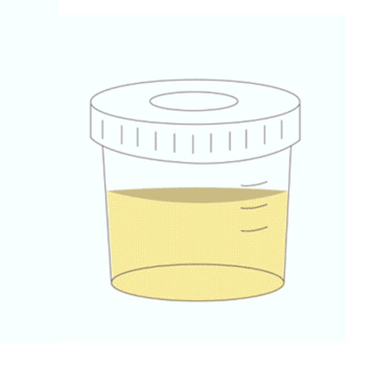
Automated peripheral blood smear and urine sediment analyser
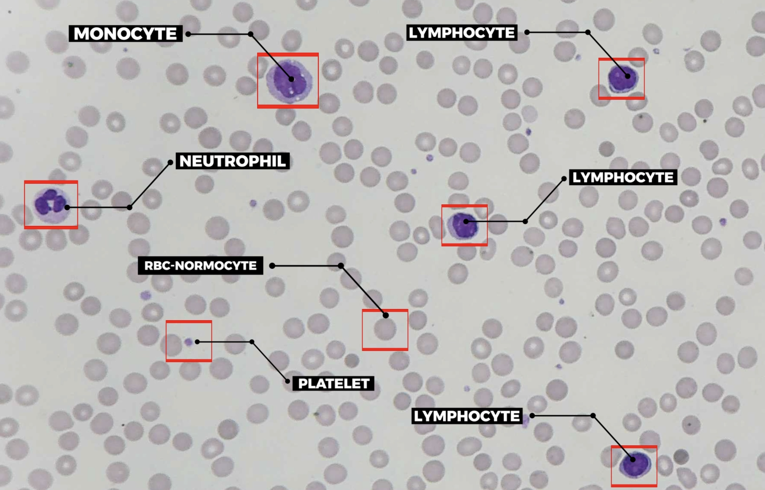
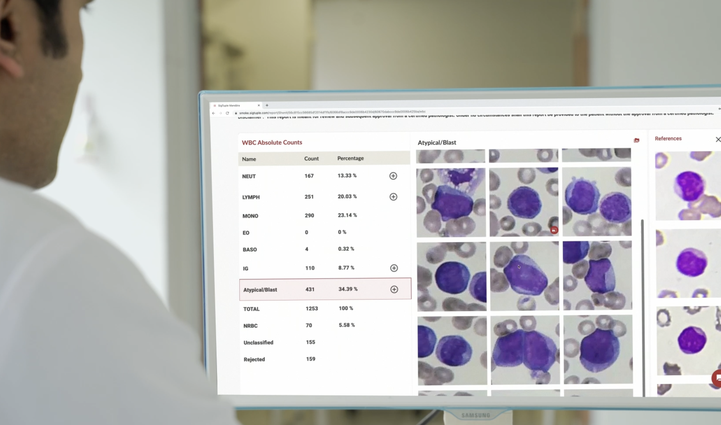
An in-vitro diagnostic device designed to automate manual microscopy in a diagnostic laboratory. It uses robotics and AI to digitize blood and urine samples on a glass slide to enable AI aided remote review
2
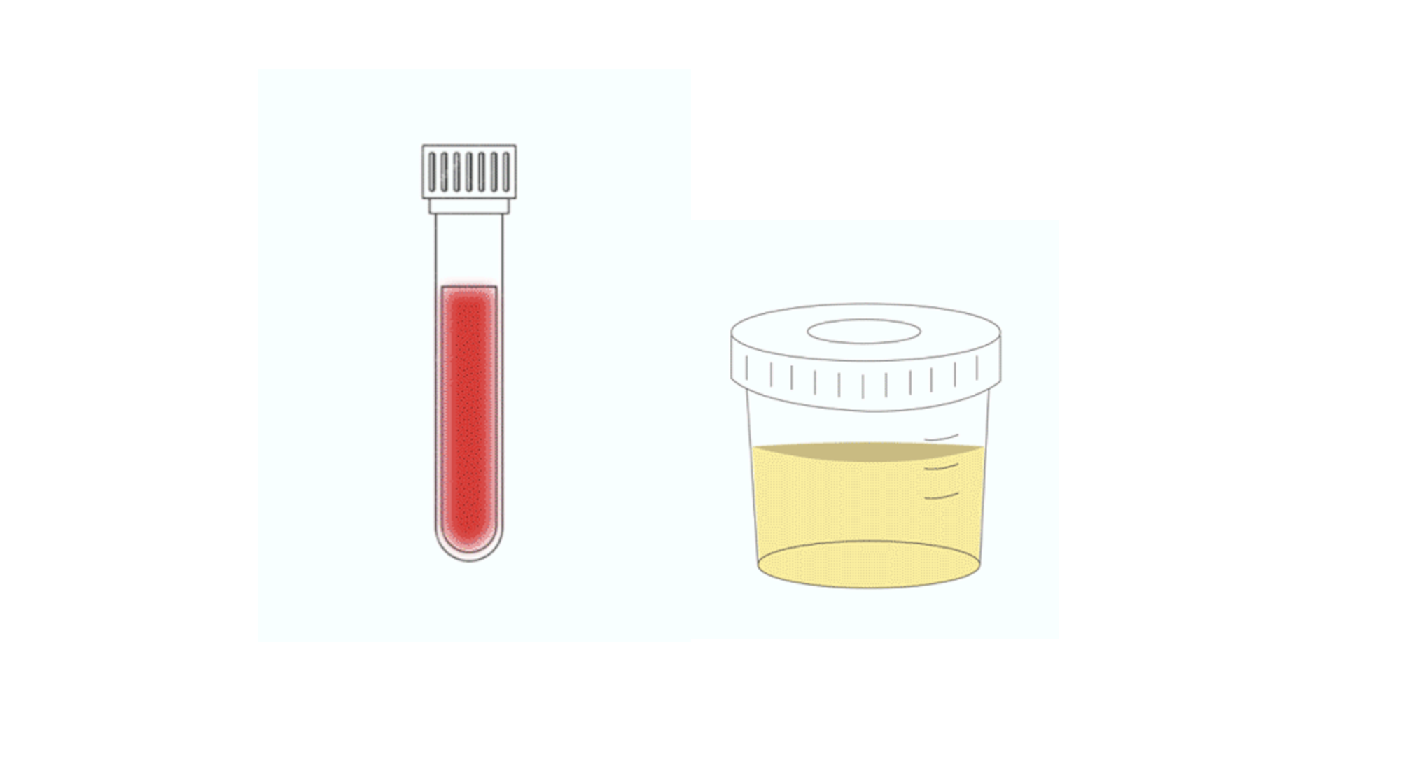
Applications
20+
Blood slides/hr
40+
Urine samples/hr
40×
Magnification

(trademark).svg)