Hero + Carousel
✓ CE Certified
AI100 Application
Shrava™

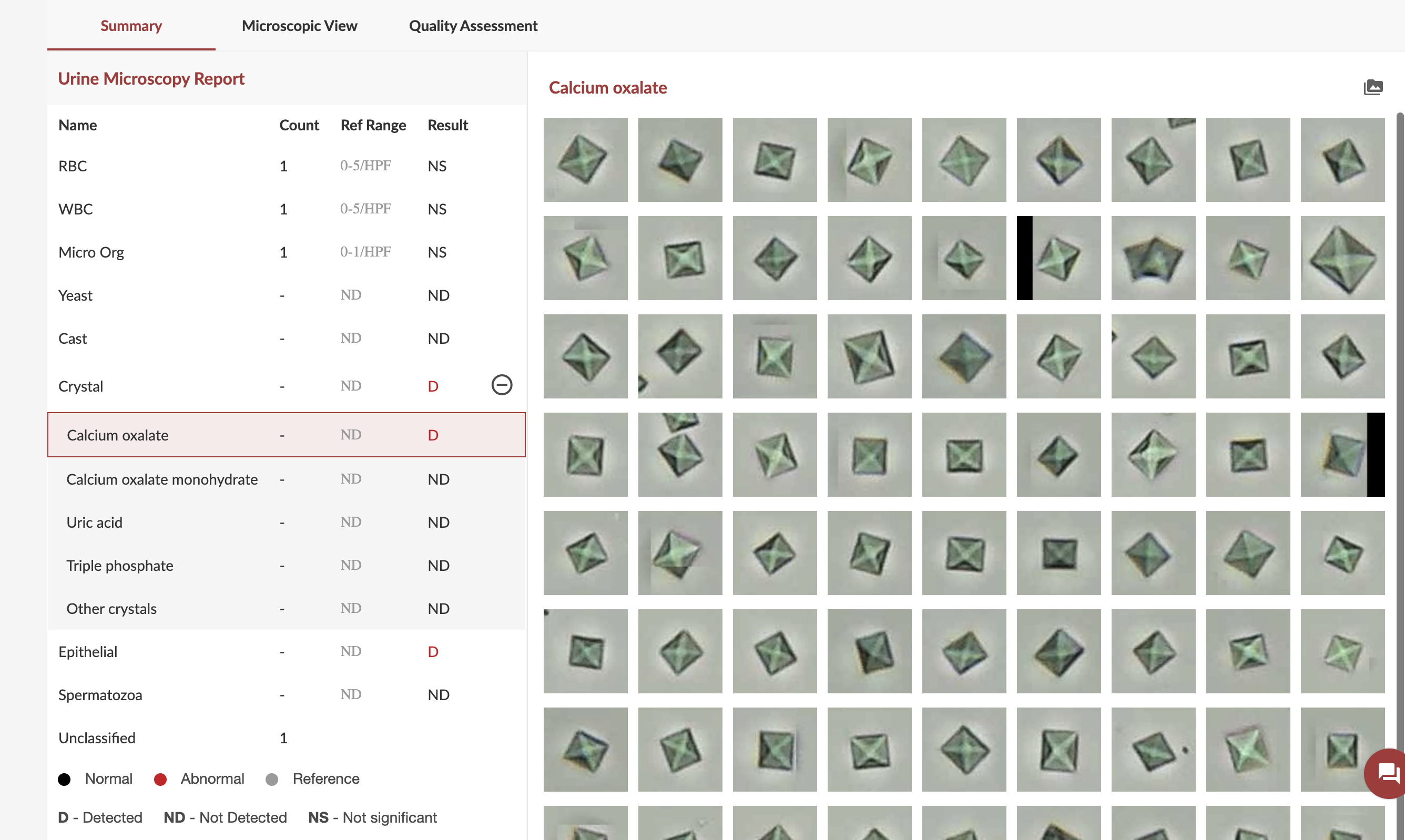
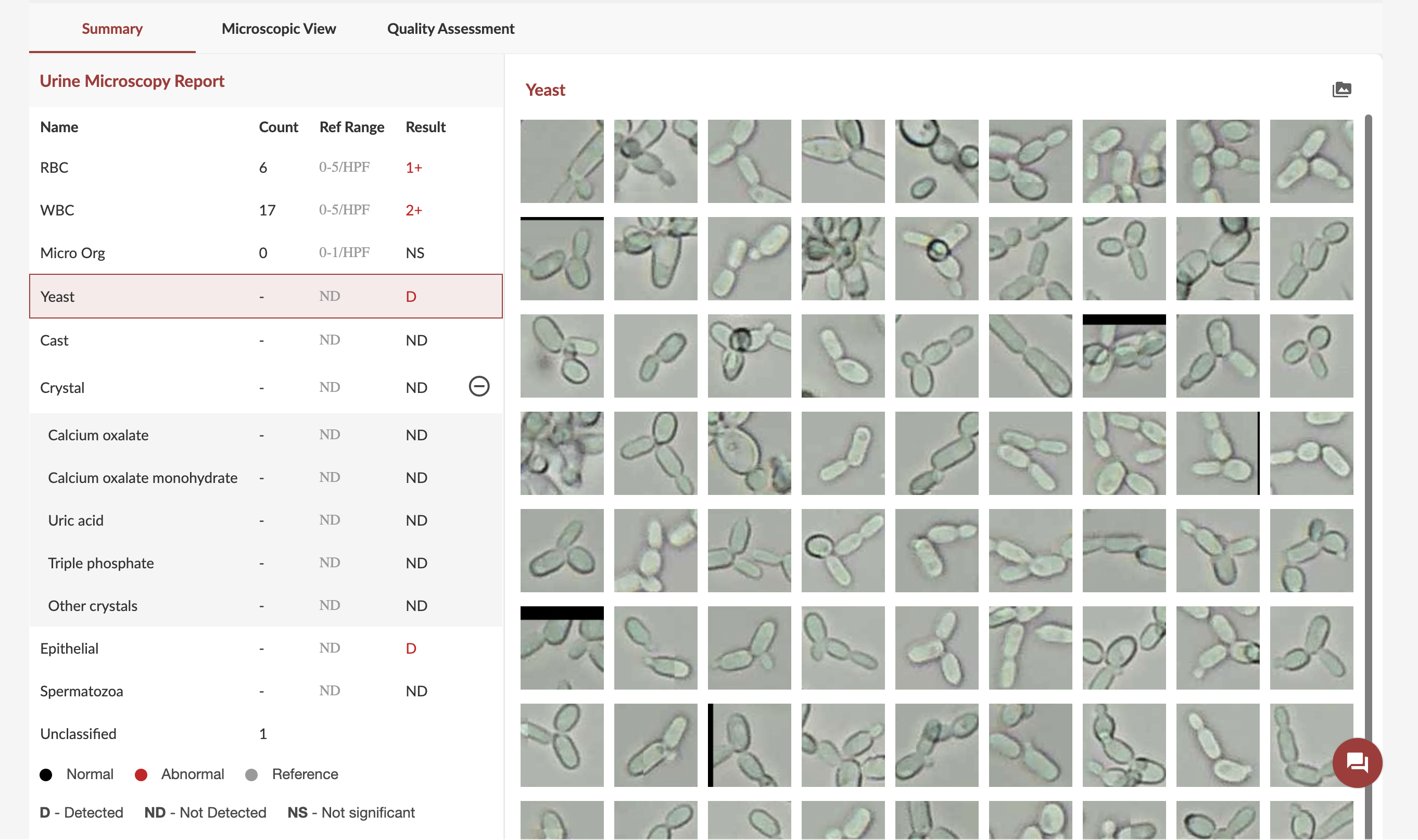
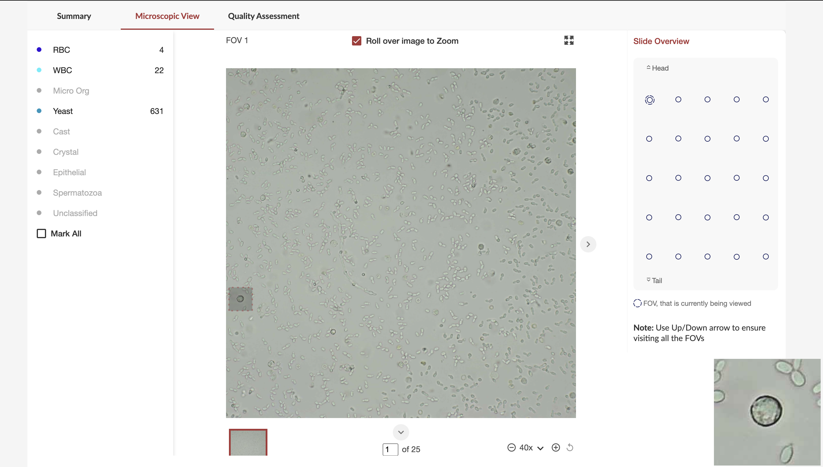
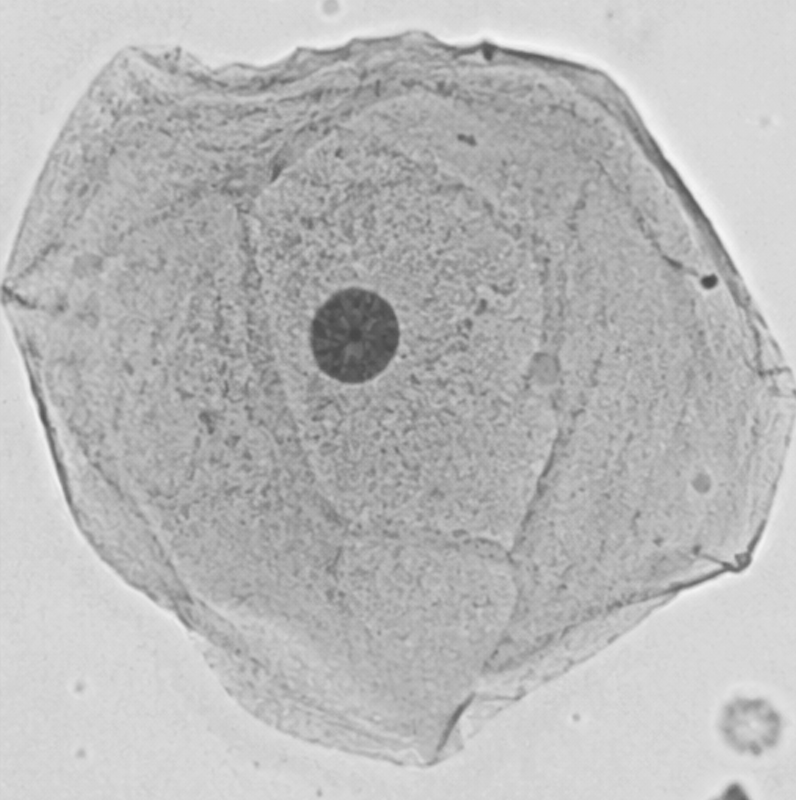
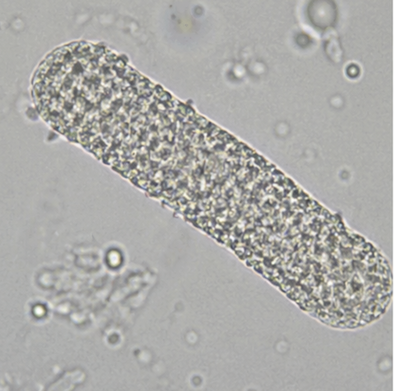
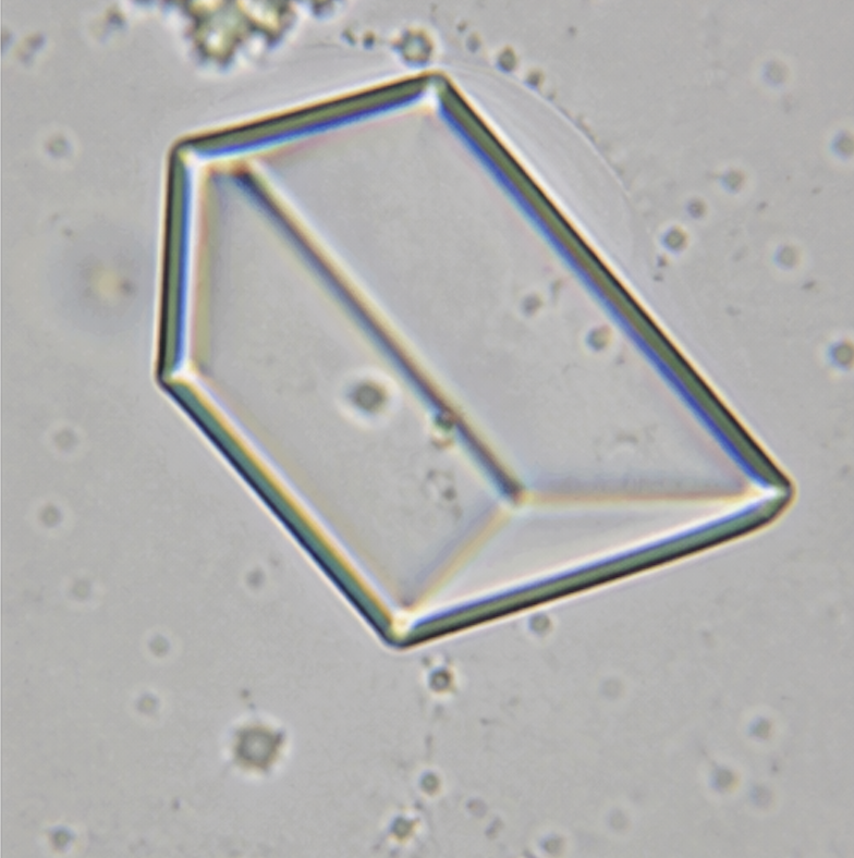
Urine Sediment Analyser

An AI application to analyse urine sediment. It identifies and pre-classifies multiple elements present in urine sediment

12
Element types
25
HPFs per sample
40+
Samples per hour
40x
Magnification
← Back to AI100
Shrava screenshot 1 Shrava screenshot 2 Shrava screenshot 3
2 / 3
Shrava— Element classification gallery
What Shrava Detects
What Shrava™ Detects

Complete urine sediment characterisation

Shrava™ analyses all major categories of urine sediment elements, providing semi-quantitative and quality results with visual evidence for pathologist confirmation.

Cells

Cells & Organisms

Automated identification and counting of all cellular elements in urine sediment — with per-high-power-field (HPF) quantification.

  • Red blood cells
  • Pus cells / WBCs
  • Epithelial cells
  • Yeast cells
  • Bacteria (bacilli & cocci)
Casts

Casts

Detection and classification of urinary casts — cylindrical structures formed in kidney tubules that indicate the location and severity of renal pathology.

Crystals

Crystals

Automated identification of crystalline structures, providing diagnostic context for metabolic disorders and infections

  • Calcium oxalate crystals
  • Uric acid crystals
  • Triple phosphate crystals
Clinical Applications

Conditions detected by Shrava™

Urine microscopy with Shrava™ supports the diagnosis and monitoring of urological, renal and metabolic conditions.

Infections
  • Urinary tract infection (UTI)
  • Bacterial infections
  • Fungal infections (Yeast)
Renal Conditions
  • Glomerulonephritis
  • Nephrotic syndrome
  • Acute tubular necrosis
Metabolic
  • Urolithiasis (kidney stones)
  • Gout (uric acid crystals)
  • Internal trauma (haematuria)
How Shrava Works
Workflow

How Shrava™ works

From urine sample to structured sediment report — fully automated and pathologist-reviewed.

1
Sample Preparation
The raw uncetrifuged urine specimen is loaded into SigTuple's proprietary Urine cartridge - GS200μ. After loading 100μL, place the cartridge on a horizontal surface for 5 minutes and then insert the cartridge in AI100
2
Automated Scanning
The AI100 robotically scans the slide across multiple fields at the appropriate magnification — capturing comprehensive, high-resolution images of the entire sediment spread systematically.
3
AI Element Detection & Classification
The captured images will be uploaded to cloud. In cloud, Shrava™'s AI models identify and classify every element in the sediment images — cells, casts, crystals and organisms — assigning type, count and clinical significance flags to each.
4
Structured Report with Visual Evidence
A structured report is generated with element counts (per HPF), visual image galleries of each category and automatic clinical flags — giving the pathologist complete visual evidence for each finding.
5
Remote Pathologist Review
The pathologist reviews and edits the report from any browser — from any location. They can zoom into any element gallery, override AI classifications and add clinical notes before final sign-off.
Call to Action

See Shrava™ in action

Book a demo and understand how our AI100 with Shrava can transform your urine testing workflow