✓ US FDA 510(k) Cleared
✓ CE Certified
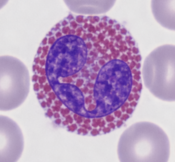
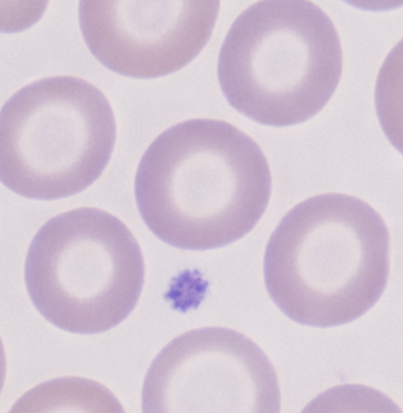
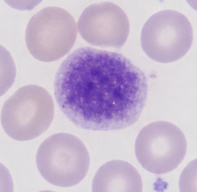
AI100 Application

Peripheral Blood Smear Analyser
An AI-powered application that automates the manual microscopic review of peripheral blood smears (PBS) — classifying every cell with visual evidence for remote pathologist sign-off.
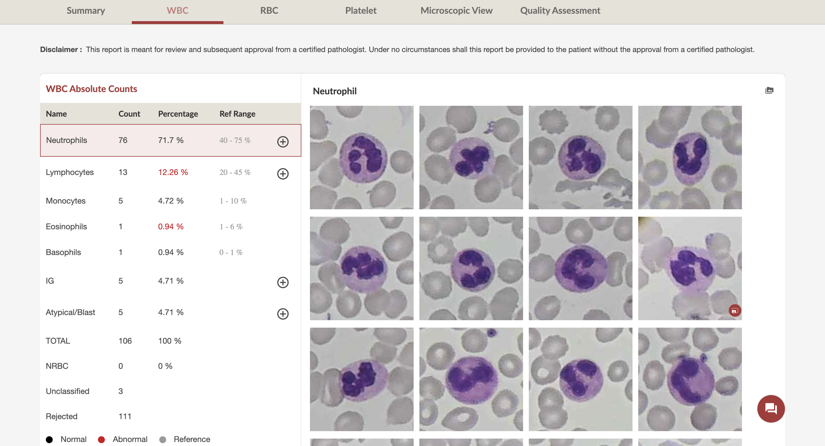
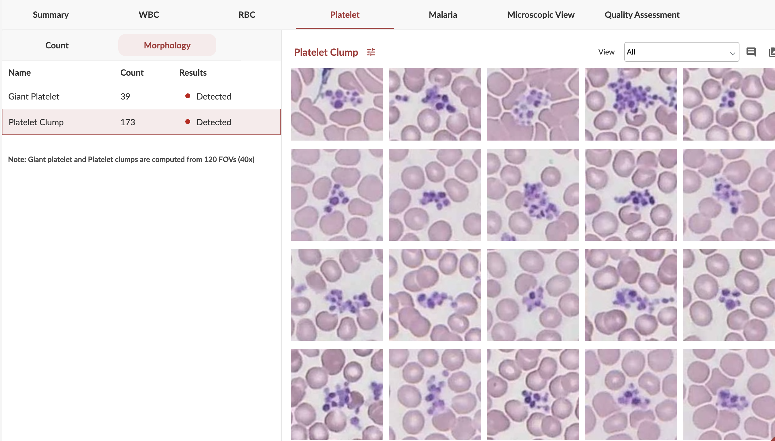
7-part
WBC differential
30+
Cell types
20
Slides per hour
40x
Magnification
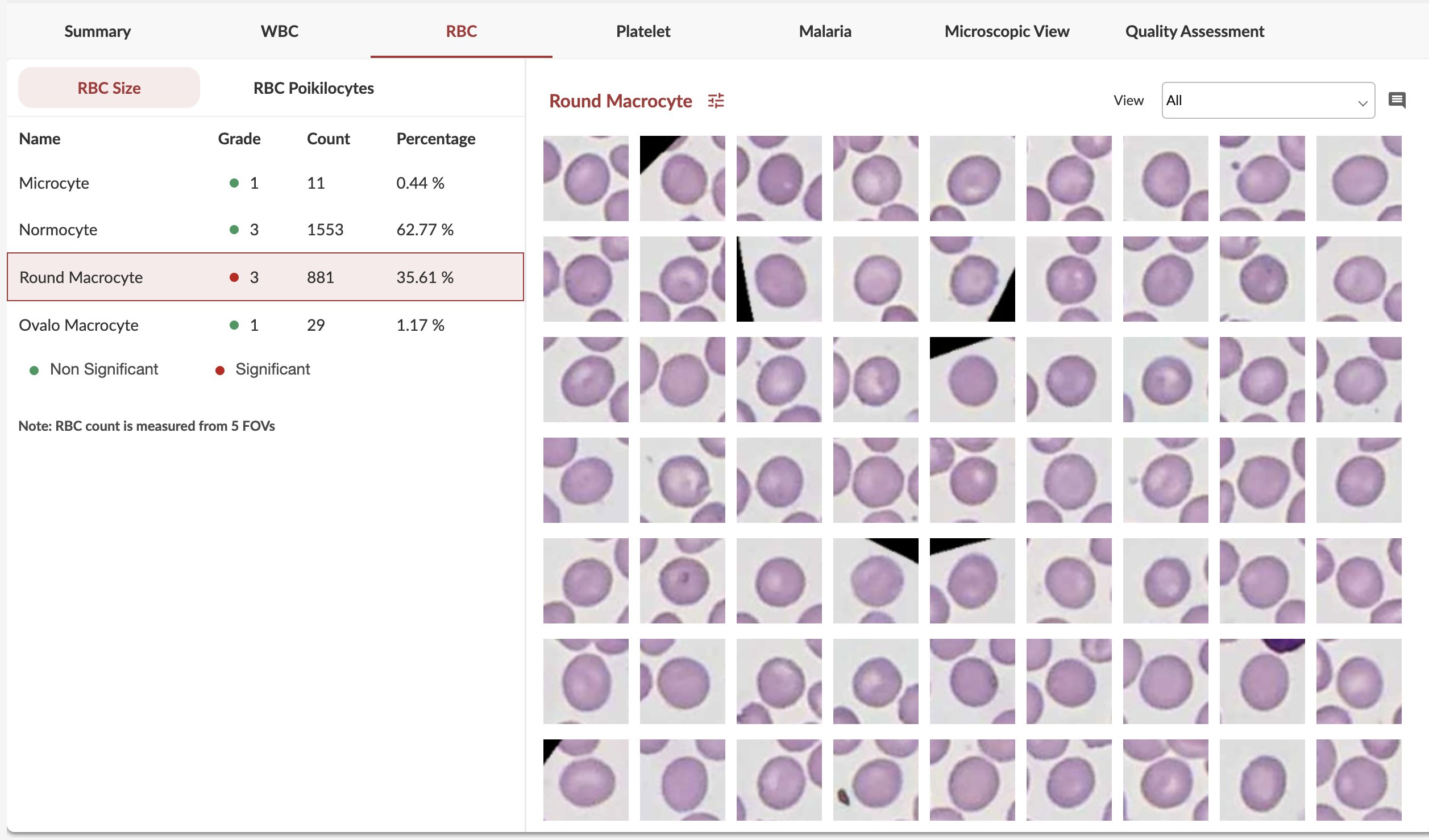
Shonit™ — RBC morphology report with ICSH grading